
Cytological tests
The shipping of biological specimens must comply with the protocols established by the courier or transport service used. In some cases, air transport requires compliance with the International Air Transport Association (IATA) regulations for dangerous goods.
In the case of superficial mass effects, fine-needle sampling (i.e., aspiration or the core/“woodpecker” extraction technique) usually has a higher diagnostic yield and is more representative of the primary lesion than samples obtained by surface impression. Impression smears are prone to contamination with debris and skin or environmental flora, or may represent only secondary inflammation or infection.
The aspirated material should be spread as a monolayer (i.e., approximately half of the cells touch but do not overlap) after application to the slide (Images of lymphocytes 1 and mesenchymal cells 2)
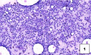
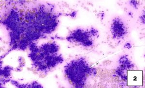
The preferred spreading method consists of a gentle horizontal spread using a second slide placed over the material after it has been deposited on the lower slide (see image 3, optimal preparation). This technique is superior to the vertical pull-apart technique (see image 4, pull-apart preparation).
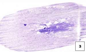
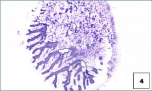
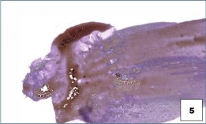
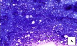
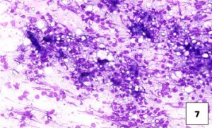
Excessive sample density and cytolysis inhibit the ability to adequately evaluate the cells in cytological samples (see slides Excessive sample density at low magnification 5 and high magnification 6; see also the slide Excessive cytolysis (cell lysis) 7 ). Only intact cells can be evaluated cytologically because cytolysis introduces artefacts that can mimic malignancy, such as nuclear enlargement and increased prominence of the nucleoli.
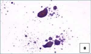
Both the pull-apart technique and the lack of effort to spread the material (i.e., simply air-drying biological material deposited as a drop) are prone to excessive sample density and cytolysis, and may be non-diagnostic (see dense sample without spreading on slide 8).
Samples with a fluid component can be spread in the same way as blood smears. Highly cellular fluids can be smeared directly; low-cellularity fluids must be centrifuged to concentrate the cells.
Slides should be kept at room temperature and protected from dust, as well as from excessive humidity and prolonged exposure to light. It is important to physically label the slides with the patient identification, as well as the origin of the cytological sample; labelling only the slide boxes or cartons is unreliable because the slides can become separated from the containers or the containers can be reused.
Samples containing an excess of ultrasound or lubricating gel may be non-diagnostic, since this material is opaque under the microscope and obscures the cells, in addition to causing poor uptake of the cell stain.
Other practical considerations for the preparation of cytological samples are to always use fresh slides, avoid the use of coverslips and acetate tape, and ensure that the cellular material is deposited towards the centre of a single side of the slide (see the sample on both sides of the slide 9).
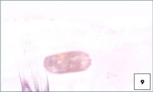
As digital slide scanning becomes widespread in diagnostic laboratories, it is not possible for a pathologist to simply turn the slide over on their microscope to focus on the material on the opposite slide. Furthermore, if the material on both sides overlaps, the net effect may be material that is too thick to evaluate adequately
Exposure of blood and cytological smears to formalin or formalin vapours (including proximity to sealed formalin jars) will interfere with adequate staining and evaluation, which may result in a non-diagnostic sample. These slide preparations should never be sent to the laboratory in the same package as formalin-fixed tissues.
Unfixed, air-dried smears are usually suitable for immunocytochemical staining (ICC); however, in some cases, it is recommended to send the samples in tubes containing transport media. If special stains, including CCI, are anticipated, the laboratory should be contacted before shipping to ensure that the specific protocol of your laboratory is followed.